

- Visibility 128 Views
- Downloads 84 Downloads
- Permissions
-
CrossMark

Integrative characterization and DNA barcoding of Fusarium isolates from diverse agroecosystems
- Author Details:
-
Monalisa Dash *
-
Nirakar Tanasingh
-
Subrat Kumar Swain
-
Kumar Krishnan
-
 Lalit Chowdhury
-
Pradipta Kumar Pati
-
Prakash Balu
-
Gopal Krishna Purohit
-
Mohammad Amin Wani
Lalit Chowdhury
Fusarium comprises diverse filamentous fungi causing economic losses through pathogenicity and mycotoxin production. Identifying Fusarium species is crucial for understanding ecology, pathogenicity, and agricultural management. This review discusses advances in characterizing Fusarium from agroecosystems, including sampling, cultural, morphological, biochemical evaluations, pathogenicity research, and molecular analysis. Collections from infected plants and rhizospheric soils reveal extensive phenotypic and genetic diversity. Traditional assessments are limited by phenotypic variability under environmental conditions. Biochemical tests enhance differentiation but often lack species-level identification. The ITS sequence is the gold standard for species identification using molecular techniques. Phylogenetic analyses of ITS and loci (e.g., TEF-1α, RPB2) reveal clusters and evolutionary history. This review emphasizes the need for a local barcode reference library combining morphological and sequence data for rapid identification in outbreaks. Pathogenicity evaluation on crops associates genotype with phenotype and informs host–pathogen interaction studies and breeding. Coordinated Fusarium monitoring and molecular surveillance with data sharing are critical for designing location-specific disease control. This integrated approach serves as a framework for understanding Fusarium biodiversity and research on plant pathogenic fungi.
1. Introduction
Fusarium species are among the most important plant pathogenic fungi, causing diseases in major crops worldwide.[1][2] These diseases include wilt, root rot, crown rot, and head blight, causing yield losses and poor grain quality in cereals, legumes, vegetables, and fruits. Fusarium-associated diseases are worsened by mycotoxins like fumonisins, trichothecenes, and zearalenone, which risk human and livestock health through contaminated food.[3][4] These impacts necessitate expanding our knowledge of Fusarium diversity and pathogenicity for developing management options. However, identifying Fusarium spp. remains challenging due to species richness and morphological plasticity. Classical identification using cultural and morphological characters is often inadequate for species delimitation, due to overlapping traits and environmental effects on phenotypic expression.[5]
The limitations of taxonomy based on morphology have been highlighted by the presence of cryptic species complexes within Fusarium, even when these cryptic species exhibit minimal morphological variation. These hidden groups of taxa have been identified in several significant pathogen groups, such as the Fusarium oxysporum species complex (FOSC) and the F. solani species complex (FSSC), emphasizing that traditional methods fail to capture the complete diversity within this genus.[6][7] Additionally, interspecies hybrids, horizontal gene transfers, and anastomoses are intricately linked to species boundaries and often complicate the process of defining species solely based on their phenotypes. Consequently, botanical pathologists and mycologists have recognized the necessity of incorporating molecular data into taxonomic practices to achieve accurate, reproducible, and universally comprehensible species identifications.[8]
Owing to the complex taxonomy involved, integrative approaches that combine phenotypic data from traditional methods, such as classical cultural and morphological characterization, with biochemical tests and molecular tools have been adopted to accurately identify Fusarium spp.[9] Molecular markers, like the internal transcribed spacer (ITS) region of ribosomal DNA and genes encoding proteins such as translation elongation factor 1 alpha (TEF 1α), RNA polymerase II subunits (RPB1 and RPB2), or β-tubulin, are frequently used to distinguish closely related species or to gain a deeper understanding of phylogenetic relationships.[10][11] The advancement of multi-locus sequence typing (MLST) and DNA barcoding schemes has significantly improved diagnostic precision, enabling researchers to identify and differentiate pathogenic lineages that are not otherwise distinguishable by their morphology.[12]
Despite inconsistent protocols for characterizing Fusarium across laboratories and regions, the lack of comprehensive barcode reference libraries with morphological, biochemical, and sequence data remains a major obstacle for comparing studies.[13] This impedes pathogen monitoring, strain identification, and resistance-breeding programs. Systematic reviews are needed to gather existing methods and suggest unified characterization strategies for widespread adoption.
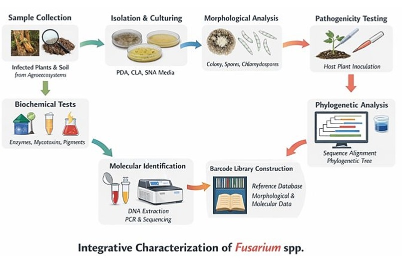
This review summarizes approaches for describing Fusarium isolates from agroecosystems. We explored morphological methods, biochemical tests, and molecular techniques, particularly DNA barcoding, for species identification. We emphasized regional barcode libraries connecting genetic signatures with phenotypic traits for identification.([Figure 1])
Isolating and identifying Fusarium species involves sampling, isolation, and morphological description. Phenotypic data includes biochemical tests, while molecular characterization involves DNA extraction, PCR, and phylogenetic analysis. Data are compiled for diagnostics.
2. Systematic Collection and Preservation
The systematic gathering and preservation of isolates from field samples are fundamental to understanding Fusarium species diversity in agricultural ecosystems.[14][15] Effective sampling requires protocols focusing on Fusarium reservoirs.[15] Infected plant materials should be collected aseptically from lesion edges. Soil cores from symptomatic plants and rhizosphere (2–4 mm soil adhering to roots) contain dense Fusarium populations. Geography and crop diversity guide sampling across varied agroecosystems with different climates, soils, and practices. Sampling should cover diverse zones and crops to determine host specificity and reveal regional pathogen ecology. Surface sterilization and selective media are essential for Fusarium isolation. Plant tissues are washed with ethanol or sodium hypochlorite before plating on PDA, CLA, or Fusarium selective media. Soil samples are plated or baited to stimulate Fusarium growth.[16]
|
Step |
Procedure / Method |
Purpose / Notes |
References |
|---|---|---|---|
|
Plant Tissue Sampling |
Collect symptomatic tissues from lesion margins using sterile tools; store in moist, cool conditions |
Ensures high pathogen load and recovery of viable Fusarium spp. |
17, 18 |
|
Soil/Rhizosphere Sampling |
Collect rhizospheric soil at 5–15 cm depth; use composite sampling near infected roots |
Targets soil-residing Fusarium propagules and increases detection sensitivity |
18, 19 |
|
Surface Sterilization |
Treat samples with 70% ethanol or 1–1.5% sodium hypochlorite for 1–2 min, followed by sterile water rinse |
Removes surface contaminants while preserving internal pathogens |
20 |
|
Isolation Media |
Plate onto PDA, CLA, or Fusarium Selective Medium (FSM); incubate at 25–28°C |
Enhances selective recovery of Fusarium colonies |
17,21 |
|
Morphological Confirmation |
Observe macroconidia, microconidia, chlamydospore structures using compound microscopy |
Confirms genus-level identity prior to molecular testing |
22 |
|
Cryopreservation |
Store culture in cryovials with 15–25% glycerol at −80°C or in liquid nitrogen |
Long-term storage with minimal genetic drift |
23 |
|
Mineral Oil Storage |
Overlay cultures on slants with sterile mineral oil and store at room temperature or 4–10°C |
Low-cost medium-term storage alternative |
24 |
|
Lyophilization |
Freeze-dry fungal cultures and seal under vacuum; store in sterile ampoules at 4°C |
Best for long-term preservation, though rehydration protocols must be optimized |
25 |
|
Metadata Documentation |
Record host species, geographic coordinates, sample type, climate data, soil type, collection date, and cultural traits |
Essential for future traceability, ecological studies, and comparative analysis |
17,26 |
Preserving isolates maintains viability and stability for future studies. Cryopreservation in liquid nitrogen or at −80°C enables long-term storage, reducing metabolic activity and genetic changes, but needs special equipment. Mineral oil preservation involves inoculating mycelia into agar plugs with oil overlay at moderate temperatures. This method keeps Fusarium viable for years but requires monitoring. Lyophilization preserves cultures through vacuum dehydration at low temperature for decades if sealed. Method selection depends on infrastructure and use.
3. Cultural and Morphological Characterization
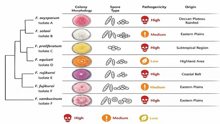
Culture and morphology-based characterization remains fundamental for Fusarium taxonomy. Isolates are cultured from infected plant tissues on nutrient media. PDA promotes mycelium growth and pigmentation between Fusarium spp.[27] Colony morphology varies in growth rate, texture, and pigmentation. CLA and SNA stimulate reproductive structures, with CLA triggering sporulation and SNA showing conidial morphology.[28] Macroconidia characteristics, including length, shape, and septation, distinguish species. Microconidia shape and formation patterns aid identification.[29] Chlamydospores help identify certain species. Cultural characterization has limitations due to phenotypic plasticity and trait overlap. Combining morphological data with molecular markers provides robust taxonomy. Morphological profiles validate molecular classifications and show phenotypic links to phylogenetic groups.
|
Species |
Colony Morphology (on PDA/CLA) |
Macroconidia |
Microconidia |
Chlamydospores |
References |
|---|---|---|---|---|---|
|
F. oxysporum |
White to purple; cottony/floccose; slow growth |
Slender, 3–5 septa, slightly curved |
Abundant, oval to ellipsoidal |
Abundant, terminal/intercalary |
|
|
F. solani |
Fast-growing; white with bluish/green center |
Broad, 3–5 septa, blunt ends |
Oval or kidney-shaped, formed in chains |
Present, terminal/intercalary |
|
|
F. proliferatum |
Pink to violet, dense aerial mycelium |
Narrow, 3–5 septa, curved |
Abundant, oval/reniform |
Rare or absent |
|
|
F. verticillioides |
Cottony white to lilac, flat colonies |
Narrow, slender, 3–5 septa, pointed ends |
Oval, produced in long chains |
Absent or very rare |
|
|
F. equiseti |
Orange-brown center, radial growth pattern |
Thick-walled, curved, 5–7 septa |
Sparse, oval |
Abundant, globose |
|
|
F. chlamydosporum |
White to pale pink, rough mycelium |
Small, slightly curved, 3–5 septa |
Sparse, ovoid |
Very abundant, diagnostic trait |
4. Biochemical and Pathogenicity Assessments
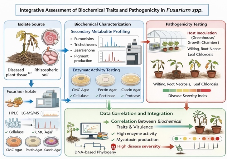
Secondary metabolite profiling distinguishes species based on metabolite production and pathogenic potential. Fusarium spp. produce mycotoxins (fumonisins, trichothecenes, zearalenone, and enniatins), which are phytotoxic and hazardous through food contamination. These metabolite profiles differ among species, serving as chemotaxonomic markers. Analytical methods (HPLC, GC MS, and LC MS/MS) detect these compounds in cultures or infected plants.[35][36] Specific secondary metabolites help distinguish related taxa that morphology cannot differentiate. Pigmentation helps help delineate Fusarium groups, with red, violet, and orange pigments being partly clade-specific. Fusarium pathogens secrete cell wall-degrading enzymes like cellulases and pectinases to break down host structures.[37] Substrate assays measure enzymatic activity, with higher activity indicating increased aggressiveness. While enzyme profiles don't define species, they help characterize virulence patterns.
Host-specific pathogenicity tests evaluate disease through seedling inoculation and symptoms. Virulence status differentiates pathogenic from opportunistic strains. Disease severity links to genetic differences, producing indices that correlate to molecular markers.[38] Biochemical profiling connects phenotypic features to virulence. Isolates with higher enzyme activity cause severe symptoms. These correlations help select markers for risk analysis. Linking metabolites to enzyme activities and pathogenicity improves understanding of Fusarium control.
5. Molecular Identification and Phylogenetics
Molecular biology enables precise identification of Fusarium species, overcoming morphological taxonomy limitations. The process involves isolating genomic DNA from fungal cultures using mechanical disruption and chemical lysis. DNA purity is verified through spectrophotometric analysis and electrophoresis. Specific primers targeting conserved regions amplify loci via PCR. The internal transcribed spacer (ITS) region serves as a barcode marker in fungal systematics, showing interspecific variation. ITS1, 5.8S rRNA gene, and ITS2 enable species distinction. ITS sequencing allows database comparison, although this locus may lack sufficient resolution within species complexes ([Table 3]). Multiple locus sequence typing provides greater phylogenetic resolution. Protein-coding genes like translation elongation factor 1 alpha (TEF-1α), RNA polymerase II subunits (RPB1 and RPB2), and β-tubulin serve as markers.[40] TEF-1α shows strong discrimination between Fusarium species, particularly within F. oxysporum species complex (FOSC) or F. solani species complex (FSSC) ([Table 2]). Multiple loci enable dataset concatenation, improving phylogenetic interpretation.
6. DNA Barcoding and Construction of Reference Library
Molecular identification and phylogeny enable precise determination of Fusarium species through DNA isolation and PCR amplification using specific primers.[41] The internal transcribed spacer (ITS) region serves as a fungal barcode locus, although it may lack sufficient resolution among Fusarium species complexes. Multilocus sequence typing (MLST) uses protein-coding genes TEF-1α, RPB1, RPB2, and β-tubulin for better phylogenetic resolution ([Table 2]), with TEF-1α showing strong species discrimination.[2][42]
Phylogenetic trees use sequence alignments as terminal nodes, analysed through Maximum Likelihood, Bayesian Inference, and Neighbour Joining methods using MEGA, RAxML, and MrBayes.[43][44] Well-supported clades link to morphological traits and pathogenic behavior. Multilocus phylogenies reveal distinct lineages with unique epidemiological profiles,[45] enabling predictive identification for disease management.
7. Disease Management and Breeding Application
DNA barcoding of Fusarium isolates aids disease management and crop breeding. Using defined isolates enhances resistance tests in breeding programs. Conventional breeding uses limited pathogen isolates, but genetic variation suggests resistance may be lost with divergent pathogen compositions. Isolates characterized through morphological and molecular examination improve resistance screening.[51] DNA barcoding helps identify virulence genes for marker-assisted selection.[41] DNA barcodes enable early pathogen detection for disease management through molecular tools that identify infections when fungicide treatments are most effective.[51][52] Surveillance systems using barcoding can be implemented through extension services. Early detection enables intervention through seed treatment or altered planting dates. Pathogen characterization enables targeted control since Fusarium populations vary regionally.[1] Control methods may not work across species due to host differences.[25][54]
8. Challenges and Future Directions
Despite advances in integrative characterization of Fusarium species, a comprehensive global species identification system has limitations. A major issue is incomplete barcode coverage for Fusarium species in public repositories.[5] Many plant disease-causing microorganisms lack high-quality sequence data with verified strains and pathogenicity, especially those underrepresented in studies and minor crops. This gap weakens molecular identification and limits phylogenetic studies. Additionally, inconsistent protocols between laboratories for cultivation, morphological documentation, and DNA sequencing reduce reproducibility and hinder data integration.[55]
|
Locus |
Target Region |
Common Primer Pairs |
Primary Application |
Reference |
|---|---|---|---|---|
|
ITS |
Internal Transcribed Spacer (ITS1–5.8S–ITS2) |
ITS1: 5′-TCCGTAGGTGAACCTGCGG-3′ ITS4: 5′-TCCTCCGCTTATTGATATGC-3′ |
Universal fungal barcode; species-level identification |
|
|
TEF-1α |
Translation Elongation Factor 1-alpha |
EF1: 5′-ATGGGTAAGGA(A/G)GACAAGAC-3′ EF2: 5′-GGA(G/A)GTACCAGT(G/C)ATCATGTT-3′ |
High-resolution species delimitation; MLST in complexes |
|
|
RPB2 |
RNA Polymerase II subunit 2 |
fRPB2-5f: 5′-GAYGAYMGWGATCAYTTYGG-3′ fRPB2-7cr: 5′-CCCATRGCTTGYTTRCCCAT-3′ |
Phylogenetic resolution in deeper clades |
|
|
β-tubulin |
β-tubulin gene region |
Bt2a: 5′-GGTAACCAAATCGGTGCTGCTTTC-3′ Bt2b: 5′-ACCCTCAGTGTAGTGACCCTTGGC-3′ |
Species-level differentiation; MLST |
|
|
RPB1 |
RNA Polymerase II subunit 1 |
RPB1-Af: 5′-CAYCCWGGYTTYATCAAGAA-3′ RPB1-Cr: 5′-CCNGCDATNTCRTTRTCCATRTA-3′ |
Complementary to TEF and RPB2 in phylogenetic studies |
Connecting traditional morphology-based identification with new methods like high-throughput sequencing (HTS) and metagenomics for non-culturable Fusarium species detection in environmental samples is challenging.[56][57] These methods complement culture-dependent techniques but need well-curated reference databases and bioinformatics pipelines. Integrating classical taxonomy with HTS data is crucial for uncovering cryptic diversity and monitoring pathogen emergence. To address these issues, developing cooperative research networks and public barcode libraries is essential.[57][58]
9. Discussion
The integrative Fusarium characterization represents a breakthrough for fungal taxonomy, plant pathology, and agricultural biosecurity. Precise diagnostics cannot rely solely on morphology due to phenotypic plasticity among cryptic species.[19][59] Biochemical, pathogenicity, and molecular data, especially ITS and TEF-1α sequencing, enable definitive identification.
Our synthesis shows variation among Fusarium populations across agroecological zones. Host selection and environmental pressures that structure diversity impact disease control.[2] Pathogenic isolates vary in virulence and toxin production, requiring connection between molecular identification and phenotypic traits. Phylogenetic analyses provide insights into evolutionary relationships, helping predict threats 8. Building DNA barcode libraries facilitates specimen identification. Challenges remain in achieving consensus taxonomy given varied methodologies. High-throughput sequencing and metagenomics will advance understanding of Fusarium biodiversity.[60][61] Harmonized data sharing and collaboration are needed to realize integrative characterization's potential for improved disease monitoring and crop protection.
10. Conclusion
Fusarium species identification and characterization are necessary to study pathogenicity, distribution, and agricultural vulnerability. This study highlights deficiencies in morphological-based methods and the need for cultural, biochemical, pathogenicity, and molecular characteristics. ITS and TEF-1α sequences with phylogenetic analysis could serve as a consistent method for identifying closely related Fusarium species. Local DNA barcode libraries linked to phenotypic and genotypic information accelerate Fusarium diagnosis without full multilocus analysis. These approaches are important for breeding programs, quarantine restrictions, and developing regional disease control strategies. Limitations include non-standardized protocols, incomplete barcode coverage, and limited high-throughput sequencing. These must be addressed through research networks, open-access platforms, and collaborations for advancement.
11. Source of Funding
None.
12. Conflict of Interest
None.
13. Ethics Approval
This research did not engage with human subjects nor animals, nor any clinical/personal/facility data. All the fungi were isolated from environmental and agricultural sources only. Therefore, ethical approval was not required for this study. All experiments were performed according to standard laboratory practices and institutional biosafety guidelines for microbial handling.
References
- Ekwomadu T, Mwanza M. Fusarium fungi pathogens, identification, adverse effects, disease management, and global food security: A review of the latest research. Agriculture. 2023;13(9). [Google Scholar] [Crossref]
- Rampersad S. Pathogenomics and management of Fusarium diseases in plants. Pathogens. 2020;9(5). [Google Scholar] [Crossref]
- Wu L, Hwang S, Strelkov SE FR, Oh SH BR. Pathogenicity, host resistance, and genetic diversity of Fusarium species from soybean in Canada. J Fungi (Basel). 2024;10(5). [Google Scholar] [Crossref]
- Todorović I MYRVJJ, Muller D. Microbial diversity in soils suppressive to Fusarium diseases. Front Plant Sci. 2023. [Google Scholar] [Crossref]
- Nikitin D, Zhelezova A, Ivanova E, Kholodov V, Ksenofontova N, Semenov M. Diversity, ecological characteristics and identification of problematic phytopathogenic Fusarium in soil: A review. Diversity. 2023;15(1). [Google Scholar] [Crossref]
- Lombard L, Sandoval-Denis M, Lamprecht S, Crous P. Epitypification of Fusarium oxysporum: clearing the taxonomic chaos. Persoonia. 2019;43(1):1-47. [Google Scholar] [Crossref]
- Chehri K, Salleh B, Zakaria L. Morphological and phylogenetic analysis of Fusarium solani species complex in Malaysia. Microb Ecol. 2014;69(3):457-71. [Google Scholar] [Crossref]
- Wang J, Liao Y, Zhang J, Li H, Ndoye M. Population structure and genetic diversity of the Fusarium graminearum species complex. Toxins (Basel). 2011;3(8):1020-37. [Google Scholar] [Crossref]
- Bugingo C, Infantino A, Okello P POPK, Turatsinze A. From morphology to multi-omics: A new age of Fusarium research. Pathogens. 2025;14(8). [Google Scholar] [Crossref]
- Schoch C, Seifert K, Huhndorf S, Robert V, Spouge J, Levesque C. Nuclear ribosomal internal transcribed spacer (ITS) region as a universal DNA barcode marker for fungi. Proc Natl Acad Sci U S A. 2012;109(16):6241-6246. [Google Scholar] [Crossref]
- Kamel T, Yasser M, Taha N, Alkhalifah D, Marhououk M, Hozzien W. Integrating morphological, pathogenic, and molecular approaches to characterize Fusarium root rot pathogens of common bean. Curr Issues Mol Biol. 2025;47(10). [Google Scholar] [Crossref]
- Mulè G GM, Hornok L, Nicholson P, Waalwijk C. Advances in molecular diagnosis of toxigenic Fusarium species: A review. Food Addit Contam. 2005;22(4):316-23. [Google Scholar] [Crossref]
- Zarrin M, Ganj F, Faramarzi S. Analysis of the rDNA internal transcribed spacer region of Fusarium species by PCR-RFLP. Biomed Rep. 2016;4(4):471-4. [Google Scholar] [Crossref]
- Ha M, Ryu H, Ju H, Choi H. Diversity and pathogenic characteristics of Fusarium species isolated from legumes. Sci Rep. 2023;13(1). [Google Scholar] [Crossref]
- Borowik P, Tkaczyk M, Pluta P, Okorski A, Stocki M, Tarakowski R. Distinguishing between Wheat Grains Infested by Four Fusarium Species by Measuring with a Low-Cost Electronic Nose. Sensors (Basel). 2024;24(13). [Google Scholar] [Crossref]
- Kim S, Kang S, Lee J. Cellulase and xylanase production by Aspergillus niger in bioreactors. Bioresour Technol. 1997;59(1):63-67. [Google Scholar] [Crossref]
- Leslie J, Summerell B. . The Fusarium Laboratory Manual. 2006. [Google Scholar]
- Nelson P, Toussoun T, Marasas W. . . 1983. [Google Scholar]
- Summerell B. Resolving Fusarium: current status of the genus. Annu Rev Phytopatho. 2019;57(1):323-339. [Google Scholar] [Crossref]
- Binetruy F, Dupraz M, Buysse M, Duron O. Surface sterilization methods impact microbial diversity. Parasit Vectors. 2019. [Google Scholar] [Crossref]
- Engalycheva I, Kozar E, Kameneva A, Sletova M, Vetrova S, Chizhik V. Molecular and morphological identification of Fusarium species causing melon wilt. J Fungi (Basel). 2025;11(12). [Google Scholar] [Crossref]
- Jia M, Fan Q, Yang Z, Wang Y, Wang X, Zeng W. Molecular and morphological evidence reveals four new Neocosmospora species from dragon trees in Yunnan Province, China. J Fungi (Basel). 2025;11(8). [Google Scholar] [Crossref]
- Whaley D, Damyar K, Witek R, Mendoza A, Alexander M, Lakey J. Cryopreservation: an overview of principles and cell-specific considerations. Cell Transplant. 2021;30. [Google Scholar] [Crossref]
- Klich M, Pitt J. A laboratory guide to common Aspergillus species and their teleomorphs. North Ryde: CSIRO Division of Food Research. 1988. [Google Scholar]
- Sharma S, Mandal S, Cramer C. Recent advances in understanding and controlling Fusarium diseases of alliums. Horticulturae. 2024;10(5). [Google Scholar] [Crossref]
- Crous P, Rong I, Wood A, Lee S, Glen H, Botha W. How many species of fungi are there at the tip of Africa?. Stud Mycol. 2006;55:13-33. [Google Scholar] [Crossref]
- Kalman B, Abraham D, Graph S, Perl-Treves R, Meller Harel Y, Degani O. Isolation and identification of Fusarium spp., the causal agents of onion (Allium cepa) basal rot in northeastern Israel. Biology (Basel). 2020;9(4). [Google Scholar] [Crossref]
- Nirmaladevi D, Venkataramana M, Srivastava R, Uppalapati S, Gupta V, Yli-Mattila T. Molecular phylogeny, pathogenicity and toxigenicity of Fusarium oxysporum f. sp. lycopersici. Sci Rep. 2016;6(1). [Google Scholar] [Crossref]
- Summerell B, Salleh B, Leslie J. A utilitarian approach to Fusarium identification. Plant Dis. 2003;87(2):117-28. [Google Scholar] [Crossref]
- Harish J, Jambhulkar P, Bajpai R, Arya M, Babele P, Chaturvedi S. Morphological characterization, pathogenicity screening, and molecular identification of Fusarium spp. isolates causing post-flowering stalk rot in maize. Front Microbiol. 2023;14. [Google Scholar] [Crossref]
- Deepa N, Sreenivasa M. Fusarium verticillioides, a globally important pathogen of agriculture and livestock: a review. J Vet Med Res. 2017;4(4). [Google Scholar]
- Xia J, Sandoval-Denis M, Crous P, Zhang X, Lombard L. Numbers to names - restyling the Fusarium incarnatum-equiseti species complex. Persoonia. 2019;43:186-221. [Google Scholar] [Crossref]
- Domsch K, Gams W, Anderson T. Compendium of soil fungi. 2nd ed. 2007. [Google Scholar]
- Parihar T, Sofi M, Rasool R, Hamid B, Bashir T, Ahmad P. Fusarium chlamydosporum, causing wilt disease of chili (Capsicum annuum L.) and brinjal (Solanum melongena L.) in Northern Himalayas: a first report. Sci Rep. 2022. [Google Scholar] [Crossref]
- Frisvad J, Andersen B, Thrane U. The use of secondary metabolite profiling in chemotaxonomy of filamentous fungi. Mycol Res. 2008. [Google Scholar] [Crossref]
- Hijaz F, Manthey J, Folimonova S, Davis C, Jones S, Reyes-De-Corcuera J. An HPLC-MS characterization of the changes in sweet orange leaf metabolite profile following infection by the bacterial pathogen Candidatus Liberibacter asiaticus. PLoS One. 2013;8(11). [Google Scholar] [Crossref]
- Perincherry L, Witaszak N, Urbaniak M WASÅ. Effects of secondary metabolites from pea on Fusarium growth and mycotoxin biosynthesis. J Fungi (Basel). 2021;7(12). [Google Scholar] [Crossref]
- Pramesh D, Prasannakumar M, Raghunandana A, Kumar M, Ngangkham U, Pushpa H. Identification and characterization of multiple fungal pathogens associated with brown spot disease of rice in India. 3 Biotech. 2024;14(8). [Google Scholar] [Crossref]
- Ma T, Yang C, Cai F, Osei R. Molecular identification and characterization of Fusarium associated with walnut branch blight disease in China. Pathogens. 2023;12(7). [Google Scholar] [Crossref]
- Alemán-Duarte MI ABGGRFBIBE. Improvement and validation of a genomic DNA extraction method for human breastmilk. Methods Protoc. 2023;6(2). [Google Scholar] [Crossref]
- Zhou L, Wu S, Ma Q, Jia X, Li P, Jin X. Phylogenomic analyses and DNA barcoding development within Moraceae: insights into genomic features, mutational hotspots, and adaptive evolution. Ecol Evol. 2025;15(11). [Google Scholar] [Crossref]
- Prahl R, Khan S, Deo R. The role of internal transcribed spacer 2 secondary structures in classifying mycoparasitic Ampelomyces. PLoS One. 2021;16(6). [Google Scholar] [Crossref]
- Zou Y, Zhang Z, Zeng Y, Hu H, Hao Y, Huang S. Common methods for phylogenetic tree construction and their implementation in R. Bioengineering (Basel). 2024;11(5). [Google Scholar] [Crossref]
- Xiang C, Gao F JI, Lei H, Hu Y, Zhang H. Using PhyloSuite for molecular phylogeny and tree-based analyses. iMeta. 2023;2(1). [Google Scholar] [Crossref]
- Bhardwaj G, Ko K, Hong Y, Zhang Z, Ho N, Chintapalli S. PHYRN: a robust method for phylogenetic analysis of highly divergent sequences. PLoS One. 2012;7(4). [Google Scholar] [Crossref]
- Iquebal M, Jaiswal S, Mishra V, Jasrotia R, Angadi U, Singh B. Fungal genomic resources for strain identification and diversity analysis of 1900 fungal species. J Fungi (Basel). 1900;7(4). [Google Scholar] [Crossref]
- Dos Reis J, Lorenzi AS dVH. Methods used for the study of endophytic fungi: a review on methodologies and challenges, and associated tips. Arch Microbiol. 2022;204(11). [Google Scholar] [Crossref]
- Wang Z, Wang Z, Lu B, Quan X, Zhao G, Zhang Z. Antagonistic potential of Trichoderma as a biocontrol agent against Sclerotinia asari. Front Microbiol. 2022. [Google Scholar] [Crossref]
- Hubka V KM. β-tubulin paralogue tubC is frequently misidentified as the benA gene in Aspergillus section Nigri taxonomy: primer specificity testing and taxonomic consequences. Persoonia. 2012;29:1-10. [Google Scholar] [Crossref]
- Liu F, Deng Y, Wang F, Jeewon R, Zeng Q, Xu X. Morphological and molecular analyses reveal two new species of Microcera (Nectriaceae, Hypocreales) associated with scale insects on walnut in China. MycoKeys. 2023;98:19-35. [Google Scholar] [Crossref]
- Kamel T, Yasser M, Taha N, Alkhalifah D, Marzouk M, Hozzien W. Integrating morphological, pathogenic, and molecular approaches to characterize Fusarium root rot pathogens of common bean in Egypt. Curr Issues Mol Biol. 2025;47(10). [Google Scholar] [Crossref]
- Stoeva D, Hristov P, Radoslavov G, Beev G, Gencheva D. Molecular identification and phylogenetic analysis of Fusarium spp. associated with barley grains using DNA barcoding in Bulgaria. Acta Microbiol Bulg. 2024;40(4):446-56. [Google Scholar] [Crossref]
- Stoeva D, Gencheva D, Radoslavov G, Hristov P, Yordanova R, Beev G. Novel DNA barcoding and multiplex PCR strategy for the molecular identification and mycotoxin gene detection of Fusarium spp. in maize from Bulgaria. Methods Protoc. 2025;8(4). [Google Scholar] [Crossref]
- Guo Q, Li S, Dong L, Su Z, Wang P, Liu X. Screening biocontrol agents for cash crop Fusarium wilt based on fusaric acid tolerance and antagonistic activity against Fusarium oxysporum. Toxins (Basel). 2023;15(6). [Google Scholar] [Crossref]
- Ahrens D. . Methods in molecular biology. 2744. [Google Scholar] [Crossref]
- Piombo E, Abdelfattah A, Droby S, Wisniewski M, Spadaro D, Schena L. Metagenomics approaches for the detection and surveillance of emerging and recurrent plant pathogens. Microorganisms. 2021;9(1). [Google Scholar]
- Wilson J, Brandon-Mong G, Gan H, Sing K. High-throughput terrestrial biodiversity assessments: mitochondrial metabarcoding, metagenomics or metatranscriptomics?. Mitochondrial DNA A DNA Mapp Seq Anal. 2019;30(1):60-7. [Google Scholar] [Crossref]
- Hilton SK CEPM, Toma I MT, Hoffman E. Metataxonomic and metagenomic approaches vs. Front Microbiol. 2016. [Google Scholar] [Crossref]
- Munkvold G. . Methods in molecular biology. 1542. [Google Scholar] [Crossref]
- Park B, Jung K, Kang S, Cheong K, Choi J, Lee Y. Cyber infrastructure for Fusarium: three integrated platforms supporting strain identification, phylogenetics, comparative genomics and knowledge sharing. Nucleic Acids Res. 2011. [Google Scholar] [Crossref]
- Chandra N, Nandini B, Wulff E, Prakash H, Udayashankar A, Niranjana S. Prospects of molecular markers in Fusarium species diversity. Appl Microbiol Biotechnol. 2011;90(5):1625-1639. [Google Scholar] [Crossref]
- Sushma S, Kolar A, Taj S, Jainab S, Tariq N, Saravanamoorthy M. Identification of antimicrobial compounds from the plant growth promoting bacteria (PGPR) tested against Fusarium wilt of tomato caused by Fusarium oxysporum f.sp. Fusarium oxysporum lycopersici J King Saud Univ Sci. 2024;36(7). [Google Scholar] [Crossref]
- 1. Introduction
- 2. Systematic Collection and Preservation
- 3. Cultural and Morphological Characterization
- 4. Biochemical and Pathogenicity Assessments
- 5. Molecular Identification and Phylogenetics
- 6. DNA Barcoding and Construction of Reference Library
- 7. Disease Management and Breeding Application
- 8. Challenges and Future Directions
- 9. Discussion
- 10. Conclusion
- 11. Source of Funding
- 12. Conflict of Interest
- 13. Ethics Approval
- References